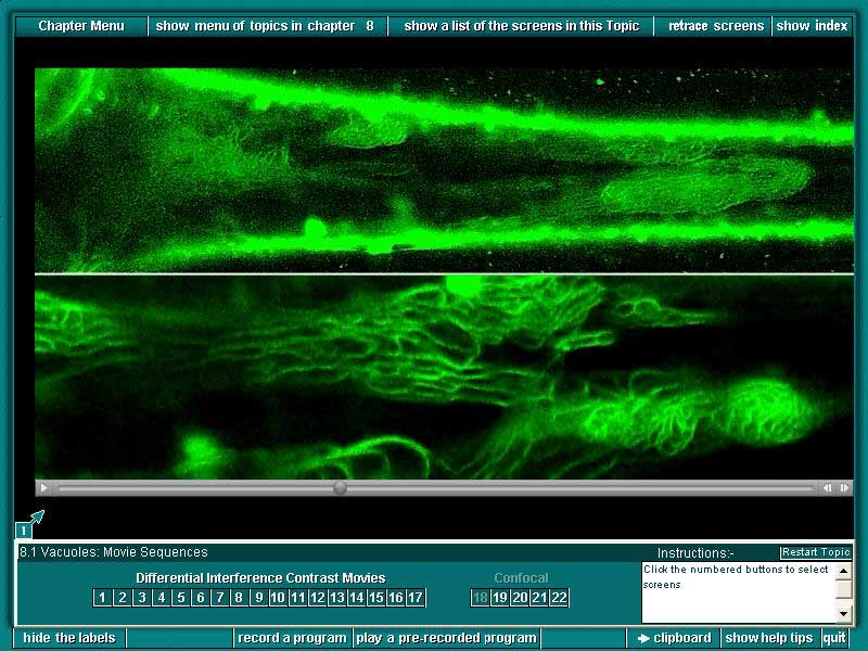
1. A tomato trichome stained with the specific fluorescent tonoplast marker MDY-64 and examined by confocal microscopy. (stills from movies) The upper panel shows a through-focus series at low magnification followed by a time lapse sequence at one plane of focus (speeded up 24x relative to real time). The lower panel is a time lapse sequence at higher magnification (same cell) to show that the channels seen in the preceding three movies are indeed delimited by tonoplast membrane (also 24x).
Previous Sample Image Next
Sample Image Back
to Plant Cell Biology on DVD Home page