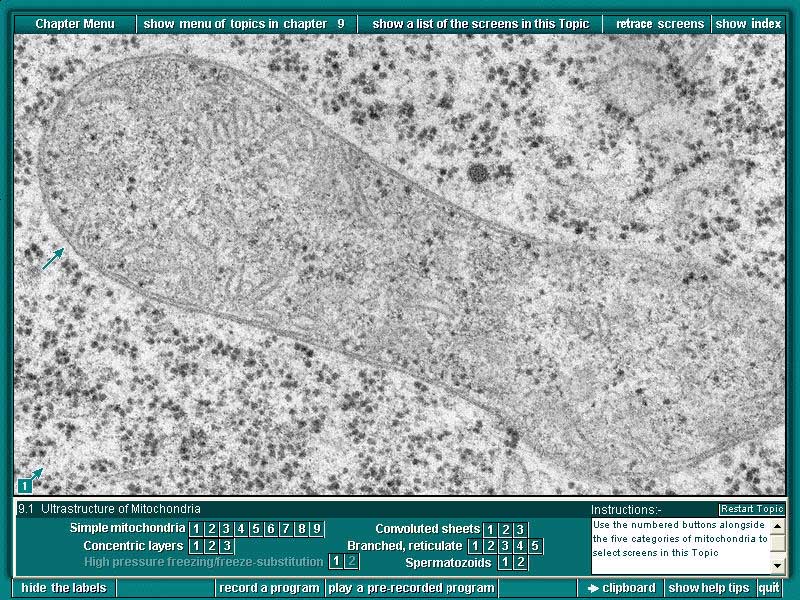
1. "Conventional" preparations for transmission electron microscopy often show a variety of artefactual distortions, though the general features are probably fairly true to life. This image comes from a superior mode of specimen preparation, namely high pressure freezing (which preserves the current state in milliseconds) followed by freeze-substitution. Note the smooth outline of the mitochondrial surface and the internal cristae. An arrow marks a connection between a crista and the inner envelope membrane. Mitochondrial ribosomes are clearly seen, and are smaller than their cytoplasmic counterparts. Eucalyptus sieberii root tip cell
Previous Sample Image Next
Sample Image Back
to Plant Cell Biology on DVD Home page