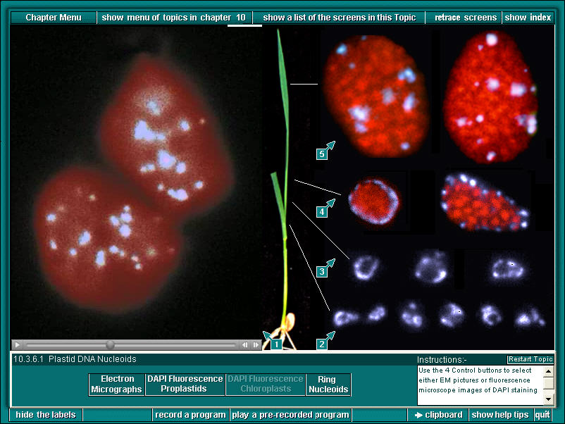
1. Through-focus images
of two spinach chloroplasts stained with DAPI to make the nucleoid DNA areas
fluorescent (there is also some background fluorescence from chlorophyll). Playing
the "movie" in effect focuses back and forth through the chloroplasts,
and shows that there are many DNA areas (nucleoids) in each one (still from
movie is shown here). There are about 20 nucleoids in each chloroplast, some
larger and more fluorescent than others, reflecting variations in DNA content.
In these mature chloroplasts the nucleoids are scattered throughout the stroms
compartment (cf. young plastids - see labels 2-4).
2. DAPI-stained proplastids from meristematic cells at the base of an oat leat.
Each of the five examples shows several nucleoids per proplastid, in no particular
arrangement.
3. DAPI-staining of proplastids in slightly older cells in the oat leaf. Now
the nucleoids are more numerous and are arranged in a ring at the periphery
of the proplastid. Chlorophyll fluorescence is not shown here (or at Label 2).
4. Two examples of young chloroplasts from cells where the oat leaf was quite
green. The red fluorescence is from chlorophyll. Two stages of proliferation
of grana are shown. In the younger stage the DNA is still in the form of a fairly
continuous ring; in the older stage it has broken up into discrete nucleoids,
but still in a peripheral ring.
5. Mature chloroplasts from the tip of the leaf. DAPI-stained nucleoids are
now scattered throughout the chloroplasts.
Previous Sample Image Next
Sample Image Back
to Plant Cell Biology on DVD Home page