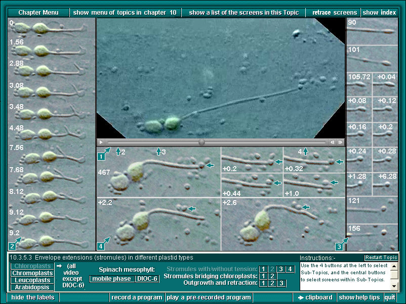
1. Stromule, 25x real time: The anchor of this stromule consists initially of a flattened apical plaque. Early in the sequence, at vertical arrow 2, a very fine strand of actin pulls out a branch from the apical plaque (see left hand panel of stills and label 2). Soon after, the plaque is transformed into an elongated vesicle, clearly still part of the stromule. At vertical arrow 3, 90 seconds into the movie, the apical vesicle transforms very rapidly (see right hand panel of stills and label 3) into a spherical body. During the next 6-7 minutes (real time) the stromule becomes extended, beaded, and at one stage has a marked "elbow" indicating a brief attachment to the cytoskeleton. The sample was moved about half way through the sequence. More stretching and contraction occurs after the move. Starting at vertical arrow 4 the stromule is released from its apical attachment and curls up into a loop, beginning and ending at the chloroplast envelope. This final process is initiated by detachment of the terminal sphere, which is released into the streaming cytoplasm (see panel of stills at lower centre and label 4). Throughout this sequence a second chloroplast is attached to the one with the stromule. The full width of the movie portion of this screen is 45 micrometres (the panels of stills are at slightly different magnifications).
2. These stills from the movie come from the region around vertical arrow 2. They portray the life history of a short-lived branch that emerged from the apical plaque of the main stromule, mover basally along the main stromule, and then retracted. The numbers are elapsed times in seconds from an arbitrary starting time (labelled zero).
3. These stills start at vertical arrow 3, about 90 seconds (real time) after the events shown in Panel 2. By then the apical plaque had transformed into an apical elongated vesicle. At time 105 (seconds) this vesicle rounds up into a spherical bead, still attached to the tip of the stromule (after 105.72 the times shown are in fractions of seconds from that moment). Although its attachment to the stromule tip seemed tenuous, the apical bead remained in place for another 6 minutes real time, while the stromule stretched and retracted many times as the chloroplast bodies were moved likes boats in a stream on the end of an anchor rope. Panel 4 (lower centre) takes over at about vertical arrow 4.
4. The terminal bead on the stromule is marked by horizontal arrows in these stills, which are from the region of the movie at vertical arrow 4. After the first still, at time 467 seconds, the times are shown as fractions of seconds elapsed from that time-point. In the space of a single second, the bead detaches from the tip and moves away in the streaming cytoplasm. It could not be identified with certainty after time point 2.6 seconds, as there were so many particles of similar appearance in the cytoplasm.
Previous Sample Image Next
Sample Image Back
to Plant Cell Biology on DVD Home page