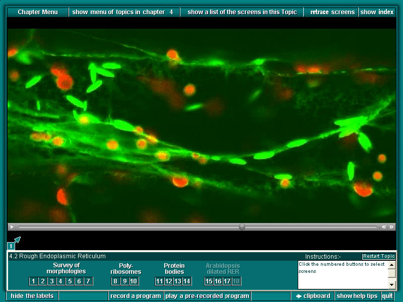
1. Confocal time lapse imaging of a hypocotyl cell in Arabidopsis that has been transformed with GFP targetted to the ER (still from movie). The brightly fluorescent spindle shapes are ER bodies; the red objects are chloroplasts showing chlorophyll autofluorescence. A nucleus is outlined by its GFP-labelled nuclear envelope. Note that many of the chloroplasts (red) have thin sheaths (green) of ER around them.
Previous Sample Image Next Sample Image Back to Plant Cell Biology on DVD Home page